 Stereotactic Radiosurgery Institute
Brain Tumor Treatment: Meningioma with Radiosurgery
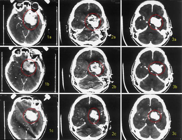
This patient had a skull base meningioma (circled in red) for which surgery (craniotomy) was performed. Because of the critical structures involved with the tumor (optic nerve, optic chiasm, carotid artery, brainstem, cavernous sinus), a large portion of the meningioma could not be removed. Radiosurgery was used for treatment of the residual tumor.
Image 1a, 1b, and 1c - The remaining tumor after the craniotomy during radiosurgery.
Image 2a, 2b, and 2c - Months after radiosurgery, the tumor (which had been growing slowly) starts to die from the inside. The tumor death, or necrosis, shows as the black spots in the tumor. The tumor is getting smaller.
Image 3a, 3b, and 3c - Months later, the body removes the necrotic or dead parts of the tumor and the meningioma is smaller. This may be only scar tissue left over afther the tumor is killed and may remain this size for the rest of the patient's life. As long as the growth of the lesion is stopped, the treatment is considered successful.
This case is presented as an example only of what has been achieved with radiosurgery by Dr. Helenowski. Each case is different and there are no guarantees that patients with similar appearing tumors will have exactly the same response even if treated the same.